Breast Health Diagnostics
Breast Cancer Resources > Breast Health Diagnostics
From Screening to Answers: How Breast Health is Assessed
Mammogram
A mammogram is a low dose x-ray of the breast used to find early signs of breast cancer. It is a quick and common imaging tool that provides your physician with clear, layered views of your breast tissue.
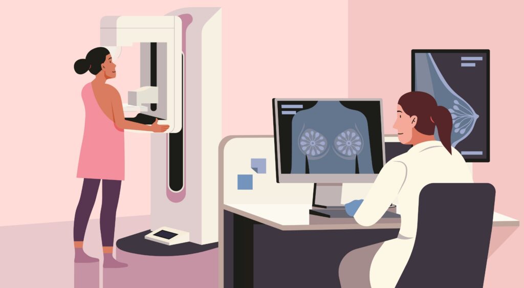
What to expect:
- You will undress from the waist up, remove any jewelry, and are given an exam robe.
- You will stand in front of the machine, and a radiology technician will position one breast at a time between two plates.
- The plates will then gently compress the breast for a few seconds to get clear images, this can sometimes cause discomfort.
- The imaging process can take about 20 minutes, after which you can resume your day.
Breast Ultrasound
A breast ultrasound is used to help your doctor see inside the breast to find the cause of lumps, pain, or abnormalities, especially in dense breasts. The ultrasound uses high-frequency sound waves to create real-time images of the breast tissue.
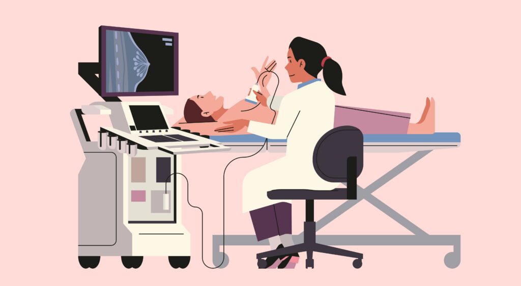
Types of Ultrasounds:
- Targeted Ultrasound: Focuses on a specific area of concern.
- Automated Whole Breast Ultrasound (AWBU): Captures images of the entire breast, offering comprehensive screening.
What to expect:
- You will undress from the waist up, removing any jewelry.
- The radiology technician will have you lie on an exam table.
- Water-based gel will be applied to your breast.
- The technologist will use a small, handheld transducer on the breast.
- This will create real-time images on a screen.
Breast MRI
A breast MRI uses magnets and radio waves to create detailed images to screen high risk women, assess cancer extent, check on implants, or evaluate dense breasts.
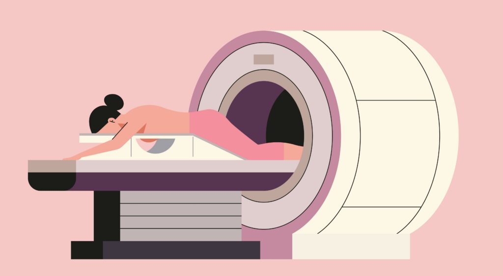
What to expect:
- You lie face-down on a padded table with openings for your breasts.
- A contrast dye (gadolinium) is usually injected into an IV to highlight abnormal tissues.
- The machine is noisy, so earplugs are provided.
- Lying still and breathing normally is crucial for clear images.
- The scan itself typically takes 30-60 minutes, with contrast given during the scan.
When something appears suspicious, imaging guides the next step, helping your care team determine whether a biopsy is needed to provide clear answers and inform the best path forward. Learn more about biopsy procedures here. https://nmcancercenter.org/breast-biopsy/